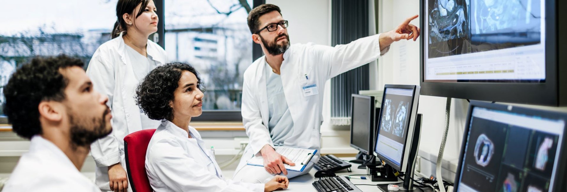
MRI in integrated diagnostics
Laboratory medicine, pathology and radiology have traditionally been fairly distinct and separate diagnostic disciplines; however, the emergence of cross-discipline technologies, such as advanced information technology, has profoundly influenced these areas and has led to the birth of new discipline, integrated diagnostics.3 This emergent field has enormous potential for revolutionizing diagnosis and for the management of diseases.3
One of these key cross-discipline technologies that has contributed to integrated diagnostics is the area of radiomics, which aims to improve tissue characterization by analyzing large numbers of quantitative features from conventional imaging studies.4 Using MRI data, radiomics can help to characterize pathologies which cannot be observed by the radiologist’s unaided eye. This data-analytics approach is expected to also improve our understanding of the underlying pathology.4 Radiomics is gradually evolving from an advanced research tool to an auxiliary clinical method, particularly in the areas of oncology and neuro-oncology, where it is expected to play an increasingly important role in the future.4 In parallel, developments in artificial intelligence, neural networks, and deep learning-based classification models have resulted in the identification of novel radiological biomarkers, which have been introduced into various aspects of clinical practice.4 Radiomic characteristics of lesions on standard-of-care imaging has already been used to generate a non-invasive biomarker for the response to immunotherapy in systemic malignancies.5 Thus, it appears these component techniques involved in integrated diagnostics are already starting to find their way into practice, or will in the near future.
In the area of breast cancer diagnosis, MRI is currently being used in virtual biopsy as MRI is a highly sensitive non-invasive diagnostic technique for characterizing breast tissue.6 MRI allows for imaging of areas that are not clearly visible on other techniques, and better highlights soft-tissue contrast resolution between lesion and adjacent breast tissue.6 The characterization of breast lesions is based on a combined assessment of morphology, internal signal variability, and enhancement pattern; all of which are best delineated on MRI.6 In the case of breast cancers, MRI can help narrow the differential diagnosis and can be used by the radiologist as a virtual biopsy tool.6
Integrated diagnostics, including blood signatures, digital pathology and radiomics may well become the most effective approach for improving the diagnosis, characterization and treatment of cancer in the near future.3 This concept may prove to be an opportunity in healthcare as it would entail better patient-centric care, improve outcomes and ultimately decrease costs over time.3 But like any new clinical technique, the potential and promise of integrated diagnostics will need to be confirmed in large-scale clinical studies.2
References
- Murray JM, Wiegand B, Hadaschik B, Herrmann K, Kleesiek J. Virtual biopsy: just an AI software or a medical procedure? J Nucl Med 2022;63:511-3.
- Chan HP, Samala RK, Hadjiiski LM, Zhou C. Deep learning in medical image analysis. Adv Exp Med Biol 2020;1213:3-21.
- Lippi G, Plebani M. Integrated diagnostics: the future of laboratory medicine? Biochem Med (Zagreb) 2020;30:010501.
- Shofty B, Artzi M, Shtrozberg S, Fanizzi C, DiMeco F, Haim O, Peleg Hason S, Ram Z, Bashat DB, Grossman R. Virtual biopsy using MRI radiomics for prediction of BRAF status in melanoma brain metastasis. Sci Rep 2020;10:6623.
- Trebeschi S, Drago SG, Birkbak NJ, Kurilova I, Cǎlin AM, Delli Pizzi A, Lalezari F, Lambregts DMJ, Rohaan MW, Parmar C, Rozeman EA, Hartemink KJ, Swanton C, Haanen J, Blank CU, Smit EF, Beets-Tan RGH, Aerts H. Predicting response to cancer immunotherapy using noninvasive radiomic biomarkers. Ann Oncol 2019;30:998-1004.
- Sharma S, Nwachukwu C, Wieseler C, Elsherif S, Letter H, Sharma S. MRI virtual biopsy of T2 hyperintense breast lesions. J Clin Imaging Sci 2021;11:18.
